To Slice or to Smear?
When the cores are being described by the sedimentologists, they need to know more than just color and visible structures. They need to know what minerals the grains are made up of, and if there are any remains of organics, such as plants and animals.
Color can give you a hint of the minerals that might be found in the core, but to be exact, we have to look more closely! There are two different ways this can be done. If the cores are mud, soft and not yet cemented into rocks, a smear slide can be made.
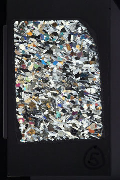
This is done by taking a tiny scraping of the sediment with a toothpick. It is put onto a microscope slide and a drop of water is added. Then it is literally smeared back and forth over the slide until there is the thinnest possible coating on the slide. It’s placed on a hotplate to dry. After the slide is dry, a coverslip is attached with optical adhesive (a glue that is invisible under the microscope so that it doesn’t change the appearance of the sediments) and the slide is left to cure. After curing under a UV lamp for a short time, it’s ready to be examined under the microscope. Then individual grains of minerals, pieces of shells, spines of sea creatures and other materials can be identified, give a much more complete picture of what the core is made of. Here is a smear slide photo from one of the 317 Cores:
If the sediments have actually been cemented into rock (lithified), then we have to use another technique. In these cases, a thin section is made. A thin section is a slice of the rock that is 30 microns thick – that’s thinner than a human hair. It’s so thin that when you hold it up you can see through parts of the slide! These are alot more time consuming to make. There are many steps, but the basic process is that a rock saw is used to cut a small rectangular block of the rock. Then that block is prepared and glued onto a slide. Then it is gradually cut thinner and thinner with a saw, and grounded down with a grinder until it is 30 microns thick. If you’d like to see the whole process – check out Making A Thin Section on our YouTube Channel. www.youtube.com/watch
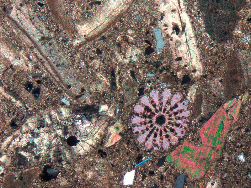
Here is a picture of what a thin section looks like under the microscope. I love looking at both types of slides and trying to identify what I see in them. It’s amazing that a rock that looks so uniform and boring can actually contain so many different things when you look closely enough! In this slide you can see microfossils quite clearly.

So hopefully, the next time you see a rock, and think “it’s just a rock”, you will remember these pictures!
