An art gallery in the middle of the South Atlantic Ocean
If nature is beautiful and science studies it, then there is beauty in science.
In each expedition the Onboard Outreach Officer (OOO) is tasked with creating content to post on social media and the website. The OOO is there to be a bridge between a scientific world and a non technical world. Coming on an expedition and staying on a ship for two months with the same people allowed for many opportunities for discussion, brainstorming or collaboration in regards to creating content.
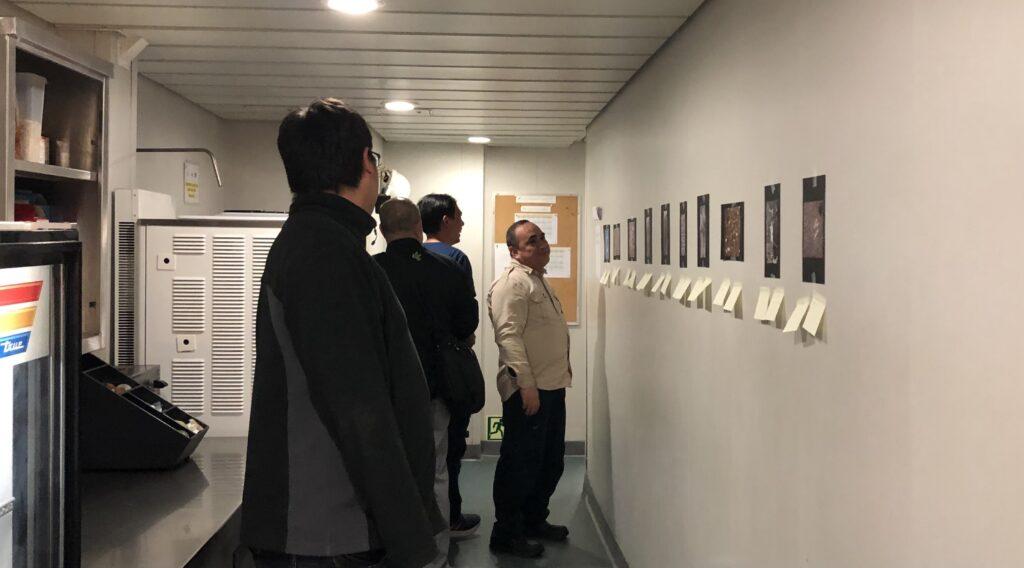
One example of such a collaboration was the Science Art Gallery aboard the JR during Expedition 393. Chiara, our very own Physical Properties specialist, correlator and stratigrapher, sent an email to the Expedition Project Manager, Trevor, and me about how she wanted to share the beautiful X-Ray images of the cores. Chiara was the spark for generating an art gallery to share with the ship crew.
Below is what Chiara had to say about her inspiration (edited for written format by Tessa):
“My inspiration came from my personal way of seeing the world and processing information. As a scientist, I have to remember a considerable amount of notions and I find it easier to recall data from my brain when they are associated with images or a silly experience. In fact, I have collected a number of silly experiences on the ship already. Since I was a kid, I enjoyed creating mind schemes with funny associations. To this day numbers have colors and sounds. I am convinced that science cannot always be dry or strictly business. Science is a wonderful universe surrounding all of us which has to be communicated to everybody. Visual communication is perhaps the easiest form of communication, a universal language which does not need grammar books. Plus, the first month on the ship we had a ship tour and I have started wondering what the ship crew (Siem and Entier staff) think about us. Do they know or understand what we do all day or do they think we are just a group of crazy people? I thought that showing them pieces of our everyday job and let them interpret freely the images would have been a good point of connection between us. Last but not least, my favorite motto is “let people surprise you”, and some of the notes are just brilliant! I would like to know the authors one by one!”
The images below that I gathered are of very thin slices of rocks (thin sections) that we can observe under the microscope and X-ray images of sediment cores.
Thin sections are aptly named. They are extremely thin (30 µm) transparent slices of rock mounted on glass microscope slides and highly polished so they can be inspected in reflected light to identify opaque minerals such as most metal oxides and sulphide minerals, for example pyrite. By looking at thin sections under a microscope with polarized light, petrologists (Petrology is the field of science that wants to understand how rocks form and change) can be more confident about their identifications of minerals and minor phases as well observe the textures of rocks. That way the cores are properly described by what alteration they have experienced or what processes have affected them. For example, the mineral olivine typically has a distinctive hexagonal shape and specific green color. However, there was a thin section that showed a mineral with hexagonal shape but filled with clay. Therefore, the petrologists knew that olivine at one point formed in the rock but now is replaced with clay.
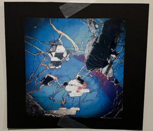
X-ray images are exactly what you think. We use an X-ray imager to scan the sediment cores to get a picture of the internal structure. Essentially, we want to know what parts are hard, soft, or voids. Using the X-ray imager provides a non-destructive way of looking inside the cores. It is similar to how we X-ray our bodies to see our bones so we don’t have to cut ourselves open. The only difference is that the X-rays for the cores are shaded oppositely than medical X-rays. What is dense is darker and what is less dense is brighter. X ray images are looked at by all of the science team but stratigraphers are the first ones to analyze the scan to estimate the quality of the core, and the layering of the sediment.
The Science Art Gallery provided two opportunities to the ship crew. The first to see what the scientists see, and then to write down what they saw when they looked at the images. It was kind of like a Rorschach test. The results were fascinating. For instance, what obviously to me was honey was snake skin to someone else or light through a canopy of leaves. It really does show that personal experiences do affect the individual’s perception.
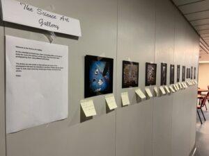
Thin Sections
What petrologists see in crossed polarized light: Plagioclase (giant gray formation at top right), and the blue-purple is clinopyroxene.
What the public saw: Marine life, Cracked tile, Broken phone screen, Blue Cheese, Pride Month
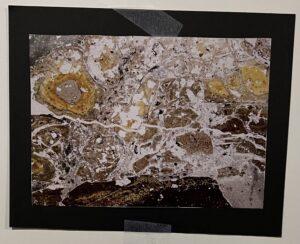
What petrologists see: The yellow blobs are altered volcanic glass in a carbonate hosted volcaniclastic sedimentary breccia
What the public saw: Aerial View from Venus, Scrambled Egg, Chernobyl, Algae, Topographic Map of a river delta and flood plain, Molds.
What petrologists see: The left side where it has a light-colored background is fresh glass, whereas the yellow background is altered glass. The dark brown spots are called varioles (from Italian for small pox) the first stages of crystallisation of the magma.
What the public saw: Leopard Seal Skin, Tad Poles, Coffee Granules, Bed Bugs, Water and oil, Tapioca Pearls.
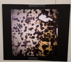
What petrologists see: A chilled margin that started with a typical basaltic composition but is now altered glass. The bright white spots are empty areas once gas bubbles called vesicles.
What the public saw: Honey, Snake Skin, Frozen Honey, Pressed flower petals, The light through the tree leaves, Gold, Thunder
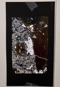
What petrologists see: A slice from a breccia with gray chunks called clasts, the black area surrounding each clast is zeolite under cross polarized light that has cemented the clasts together while the white region is carbonate that filled up a void after the clasts were cemented together.
What the public saw: Pangea starting to break, Ants vs the World, Gold Dust, Kaleidoscope, Google maps aerial view, west coast of South Africa.
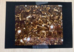
What petrologists see: Typical basalt made of a groundmass of tiny crystals. The large grey-white to black grain is a phenocryst that must have grown in a magma chamber in the crust before the magma was erupted onto the seafloor. The black spot is a vesicle where a gas bubble was trapped when the lava cooled and only later did the gas leave to make it an empty spot.
What the public saw: Stars, Black Hole, Wood stock concert, Land fill, Confetti, Interdimensional Space
X-ray images
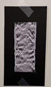
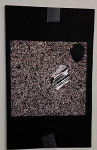
What the stratigraphers see: This core experienced high drilling disturbance because there seems to be curved lines. We also can tell this core was extremely wet since the white spots are bubbles of water.
What the public saw: Soup of the day, Snow, Powdered Milk, Moon Craters, Bone Marrow, Thick and Lengthy Muscle.
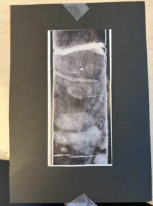
What stratigraphers see: Bioturbation in the sediment. Burrows were created by living organisms moving about the sediment and the burrows got fossilized.
What the public saw: An Ultrasound, A UFO, Worm and apple, Boulder in a desert.
When the gallery was put up it was great to watch anyone who passed, slow their pace to see what was on the usually empty wall. People from different crews started to linger and interact with each other as they interpreted the images. They got to experience science without the very common imposter feeling of not being the expert so why look at the object or talk about it. They brought themselves to the science without any judgement.