This entry is written by Debbie Thomas, co-chief scientist of Expedition 378. It comes from her Expedition 378 Odyssey blog, which can be found here.
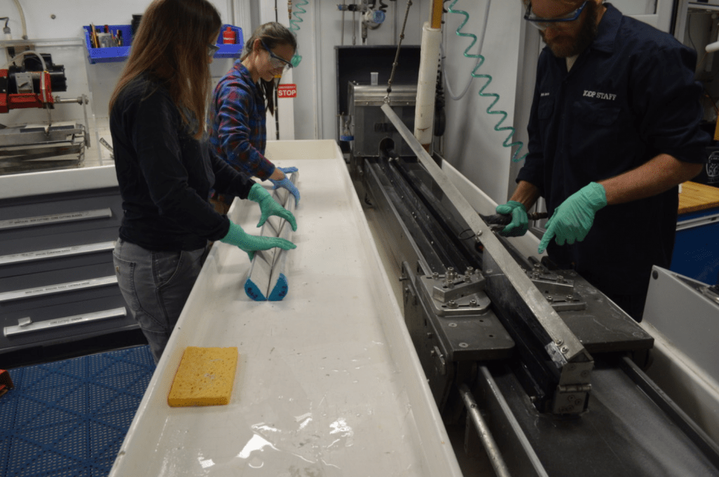
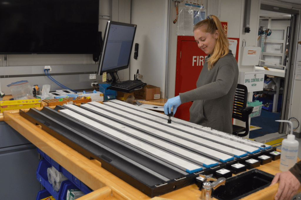
Each whole-round core section is divided lengthwise into two halves – the working half, from which discrete samples are removed for analyses of all sorts, and the archive half, which is intended for an intact legacy record for future reference. The splitting room features some very clever innovations to split soft sediment and hard rock cores:
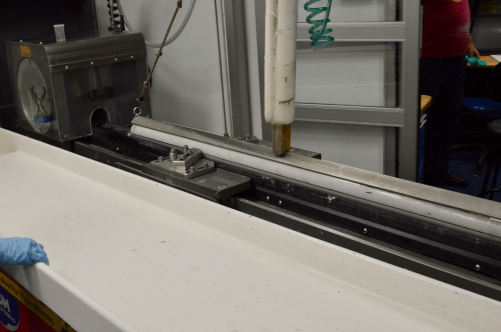
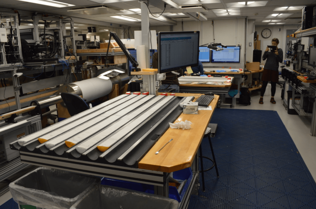
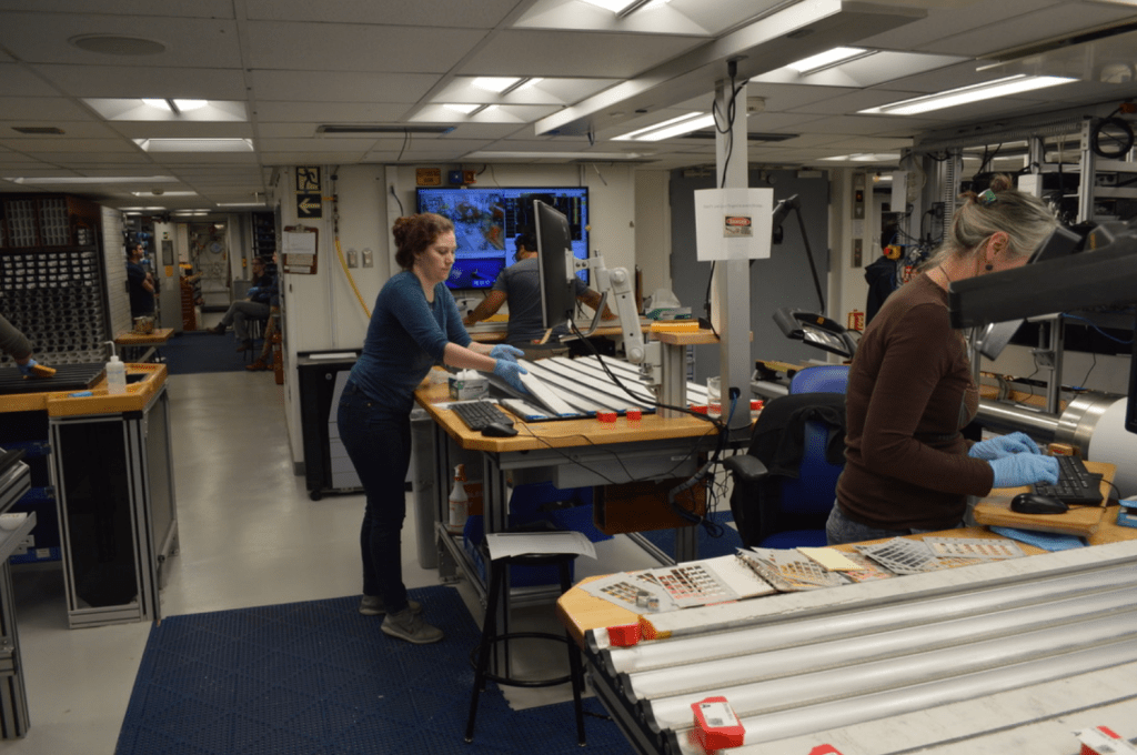
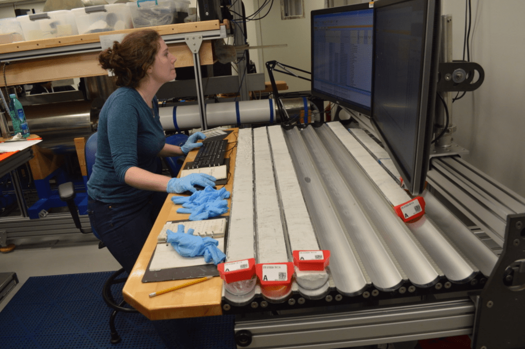
The core section is held in place by the triangular metal guide and brace on top, and a trolley just to the left of the brace pulls a piano wire through the core to slice it in half along the black line.Once split, Nicolette and Susan each take one of the ends, tap the section firmly on the table, and split the core apart, while Aaron starts cleaning the splitter:Susan took the archive half to the description table and Nicolette took the working half to the sampling table.The first stop for the archive half is the first core description table, where the split halves await their turn in the digital imager, and then the color reflectance scanner. Measuring these properties shortly after splitting is critical to capture and record the original color and composition before the sediments begin reacting in the presence of oxygen, light and warmer temperatures.It looks like Claire had the same idea. The digital imager and color reflectance tracks are to Claire’s left.Next stop is the visual description tables. Ingrid is focusing on documenting the original color of the different sediment features (while Laura escorts the sections to the scanners):Good grief, did the Munsell color chart explode on the table or did Ingrid hurl it in frustration at the difficulty of identifying the precise white of our cores?At the final description table, Laura receives the cores from Ingrid and identifies the structures and features apparent in the cores such as burrows or unique layers:
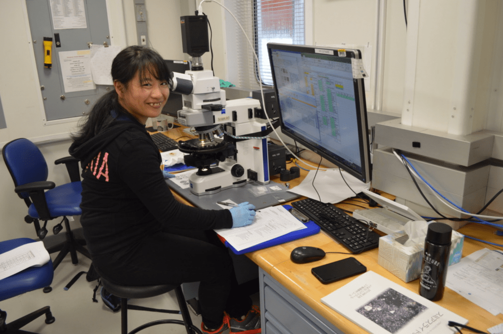
And to round out the sediment description team’s activities, Erika is assessing the sediment composition by swiping a toothpick’s worth of the sediment onto a microscope slide and sealing it with optical adhesive and a cover slip. She has the important task of estimating the relative abundance of the major types of grains that we find in the sediment – microfossils, detrital grains such as dust particles, volcanic glass, mineral grains, and any other minor phases.
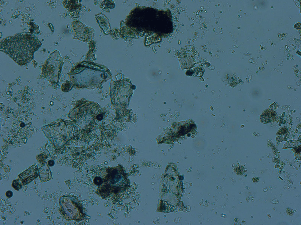
Here is an example of what she might see under the microscope from one of the JR’s library of teaching slides:
The black scale bar is at the bottom. Thank you for sharing this, Erika!We are taking a few samples from the working half of the core during the cruise to help us characterize the composition, paleomagnetic character, and physical properties of the material. Standard analyses such as moisture and density (MAD), carbonate (CARB), x-ray diffraction (XRD), paleomagnetism (PMAG) among a few others are carefully taken from the sections at the sampling table and then the sections are wrapped and slid into containers for storage in the reefer until they are shipped to the Gulf Coast Repository at Texas A&M University.
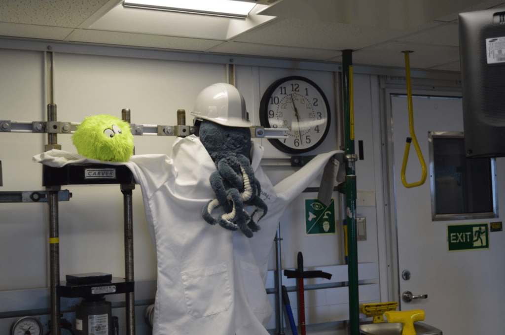
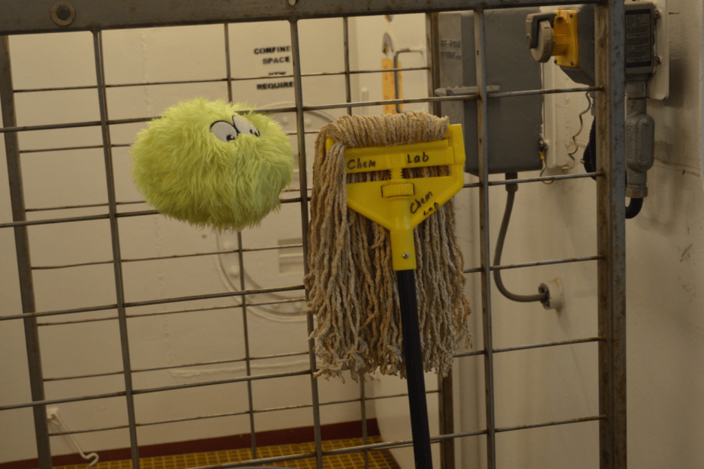
Gabby taking a physical properties measurement, and if you look closely you can see the tops of a few styrofoam plugs in the sections marking the locations of samples already removed for shipboard analysis.In other shipboard news, LC has made some pretty cool friends:
LC was quite taken with Eleni’s Octo dressed as Chang in the chem lab.