Science Art Gallery: X Ray Image 2
X ray imaging used to be run through the X Ray Image Logger where the whole round and section half cores would be run through the logger in one orientation. The result was a 2-d radiograph of any internal structures. However, the JR recently switched over to the X-Ray Linescan Image Logger (XSCAN). The bonus features is that it can not only produce high resolution images of the whole round and section half cores, but it can rotate 180 degrees around the core imaging the internal structures at multiple angles. This then allows us to see the internal structures or debris or features from more than just one 2-D view, but multiple.
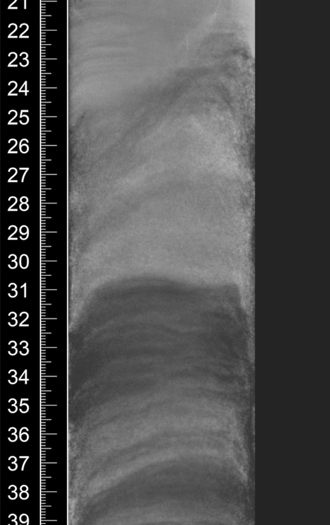
The X-ray images produced by either equipment visualize densities of the features. The lighter the white the less dense the object is, while the darker it is the more dense it is. Empty space would be completely white, while a hard rock would be the darkest thing in the image.
The X-SCAN image below comes from a sediment core. Scientifically we can see it is a sediment core because of the lighter color greys, and the layering that is evident. The curvedness of the layering near the top indicate some kind of inconsistent sedimentation.
But what do you see? Please leave a comment letting us know what you see in the image or leave a comment letting us know what it reminds you of.
Click here to return to the Science Art Gallery.